Garden Transmissions
View publication
Elisabeth Ravaud is doctor of medicine, specialised in radiology and scientific imaging and has defended a PhD in Art history. She has been working at the 'Centre de Recherche et de Restauration des Musées de France' (C2RMF) for more than twenty years, in charge of studies and research of easel paintings. She works more particularly on the contribution of scientific imaging to the knowledge and conservation of European easel paintings from the 11th to the early 20th century. Elisabetyh Ravaud also coordinated the ICOM-CC Painting Group from 2014 to 2020.
The ‘Centre de Recherche et de Restauration des Musées de France’ (C2RMF)
The C2RMF is a Department with national competence of the Ministry of Culture. It has two main missions: scientific research, centred on the knowledge of artworks from French museums in particular their original materials, and conservation/restoration of these works, carried out in its conservation studios in Paris and Versailles or by scientific support on site. Its particularity is to bring together in the same place, around the works, the combined views and skills of art historians, physicists, chemists, curators, archaeologists, restorers, photographers, documentalists, administrators and technicians. The C2RMF is aimed at the 1220 labelled "museums of France". It benefits from the support of the Ministry of Research, thanks to collaborations with the National Centre for Scientific Research (CNRS), as well as from the European Community through transnational projects.
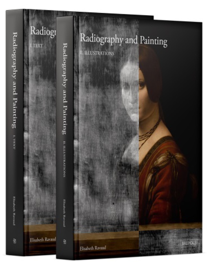
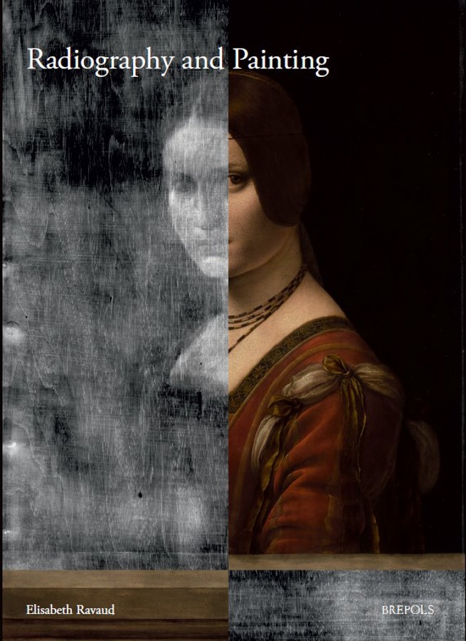
Radiography is a technique that has been employed in the study of paintings for more than a century. The history of this method of analysis indicates that its development has been modest since the 1960s, as its use has been limited to reductive approaches that take into account no more than the immediately intelligible signs. By systematically considering the physical mechanisms involved in the creation of an image, this volume seeks to demonstrate that we can access new fields of radiological analysis by identifying two categories of 'signs': those that may be obvious but whose meaning is misleading, and those which are not immediately comprehensible.
This study has been primarily based on a thorough and essential reviewing of current literature concerning the materials and processes used for the making of paintings. The semiological analysis is based on the understanding of the physical phenomena occurring in the formation of the image, and on correlations between the radiographic images of a painting and the information stemming from its observation, other scientific results, and the restoration reports. Furthermore, a number of experiments were conducted to consolidate certain assumptions regarding image-formation mechanisms. Ultimately, this book hopes to show how data resulting from radiographic analysis can be seen and set in a broader context of information on a specific work or a group of works, in order to enrich our knowledge of art history, history of technology, and conservation as well as restoration.
La radiographie est une technique appliquée aux peintures depuis près d'un siècle. L'histoire de cette technique d'analyse montre que son développement s'est trouvé limité depuis les années 1960 en raison d'une approche uniquement basée sur la prise en compte de signes immédiatement suggestifs. Or ce mode d'analyse ne permet pas d'expliquer l'ensemble des images observées sur une radiographie de peinture. En considérant de manière systématique le mécanisme de formation de l'image, ce travail souhaite démontrer que l'on peut accéder à de nouveaux champs de l'analyse radiologique en identifiant deux autres catégories de signes: les signes suggestifs mais dont la signification est trompeuse et les signes non immédiatement intelligibles.
Cette analyse a été possible grâce à une revue de la littérature la plus complète possible sur les matériaux constitutifs et les procédés employés pour la réalisation des peintures. L'analyse sémiologique est basée sur la compréhension des phénomènes physiques en jeu dans la formation de l'image et sur des corrélations entre les images observées et les informations provenant de l'observation, d'autres examens réalisés sur l'oeuvre ou encore de rapports de restauration. De plus une expérimentation a été conduite pour conforter certaines hypothèses de mécanisme de formation de l'image. Enfin, les données issues de la radiographie peuvent être replacées dans un contexte plus large de données ou informations connues sur une œuvre ou groupe d'oeuvres pour enrichir nos connaissances en histoire de l'art, histoire des techniques et conservation-restauration.
VOLUME ONE - TEXT
Preface
Acknowledgements
Introduction
Chapter I. A history of the radiography of paintings
Chapter II. The paint layer
Chapter III. The preparatory phases
Chapter IV. Supports
Chapter V. Damage and Restorations
Chapter VI. Contribution of radiography to the history of art, the history of painting practices and conservation
Conclusion
Annex 1: List of the x-rays done by Alan Borroughs during his stay in Paris in 1926
Bibliography
Index
VOLUME TWO - ILLUSTRATIONS